


Prof. Dr. Thomas Kuner
Dept. Functional Neuroanatomy, Institute for Anatomy and Cell Biology, Heidelberg University
Dr. Wolfgang Weber-Fahr
Dept. Neuroimaging, Central Institute of Mental Health, Medical Faculty Mannheim, Heidelberg University
Multiscale analysis of structural plasticity in cortical circuits
Background and past work:
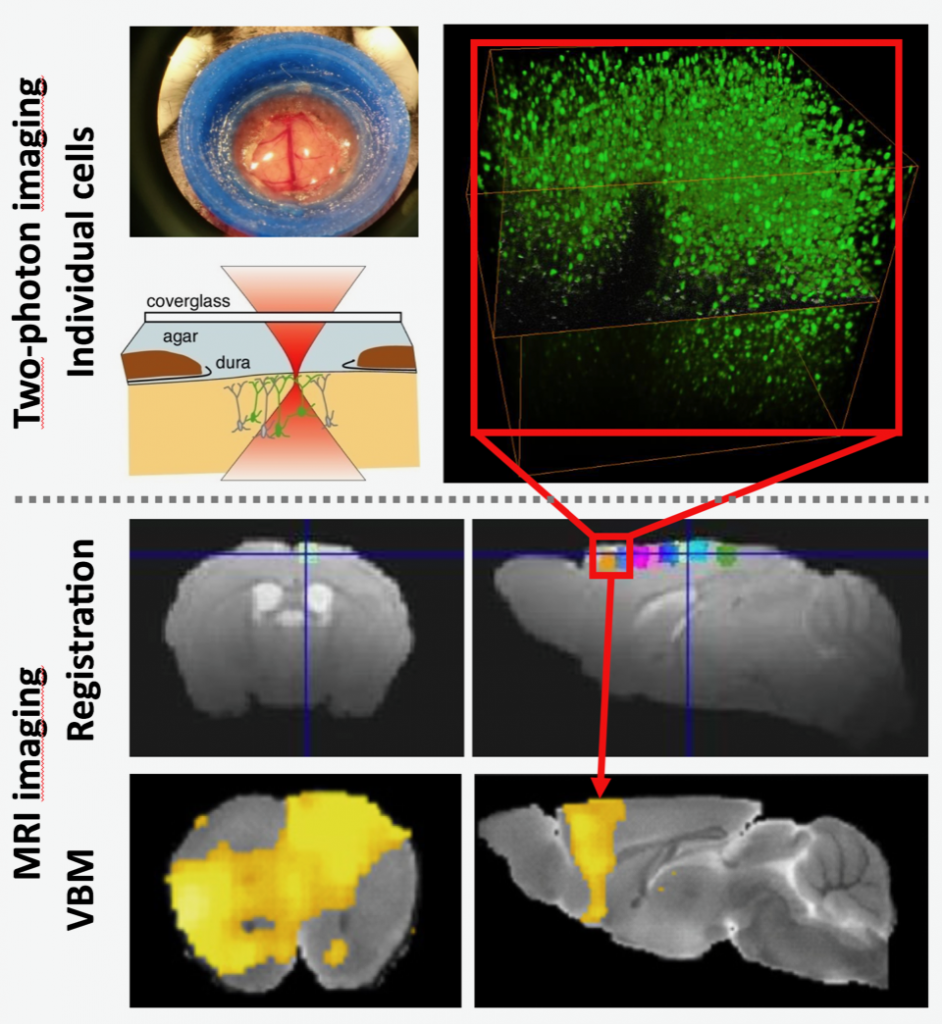
Grey matter volume (GMV) changes during chronic pain states have initially been described in humans and are believed to be relevant to chronic pain, particularly given their reversibility upon treatment. They have recently been recapitulated in rodent models, including our own meso-scale analysis of changes in grey matter volume, connectivity and functional coupling in magnetic resonance imaging (MRI) studies on mice with neuropathic pain. However, the physical underpinnings and cellular determinants of grey matter volume changes in general, and in chronic pain states in particular, has remained elusive.
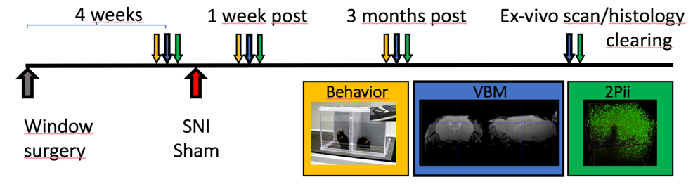
Yet, long-lasting structural changes in neocortical organization may represent a central mechanism of pain chronicity. Therefore, knowing the cellular basis of grey matter volume changes will be a major step in understanding chronic pain. During the previous funding period, we focused on subcellular changes of neuronal morphology in a mouse model of chronic neuropathic pain. In neurons of the cingulate cortex, we found changes in dendritic spine density and axonal bouton density developing on distinct timescales.
Yet, long-lasting structural changes in neocortical organization may represent a central mechanism of pain chronicity. Therefore, knowing the cellular basis of grey matter volume changes will be a major step in understanding chronic pain. During the previous funding period, we focused on subcellular changes of neuronal morphology in a mouse model of chronic neuropathic pain. In neurons of the cingulate cortex, we found changes in dendritic spine density and axonal bouton density developing on distinct timescales.
Results obtained in previous funding period:
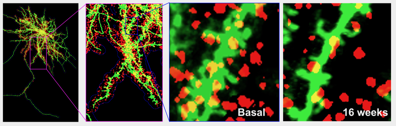 Individual cingulate neurons were imaged and changes in dendritic spines (green) and presynaptic thalamic boutons (red) were monitored over time. This example shows a decrease of both spine density and number of presynaptic boutons in response to spared nerve injury.